
Formation of Coloured Ions
Quick Notes
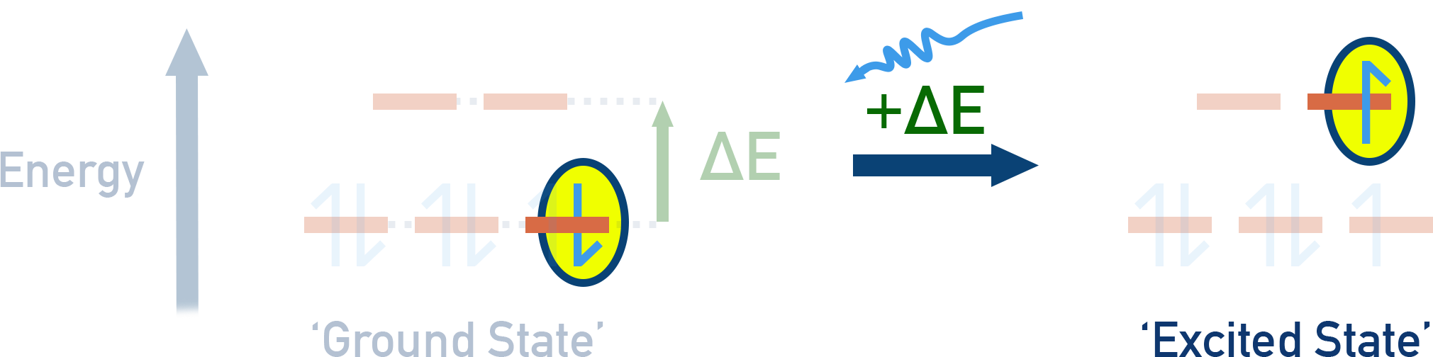
- Transition metal ions are coloured because electrons in the outer d sub-shell absorb energy from visible light and move to a higher energy (excited state).
- Energy difference (ΔE) between d-orbitals is given by the equation:
ΔE = hν = hc/λ- h = Planck’s constant
- ν = frequency of light absorbed
- λ = wavelength of light absorbed
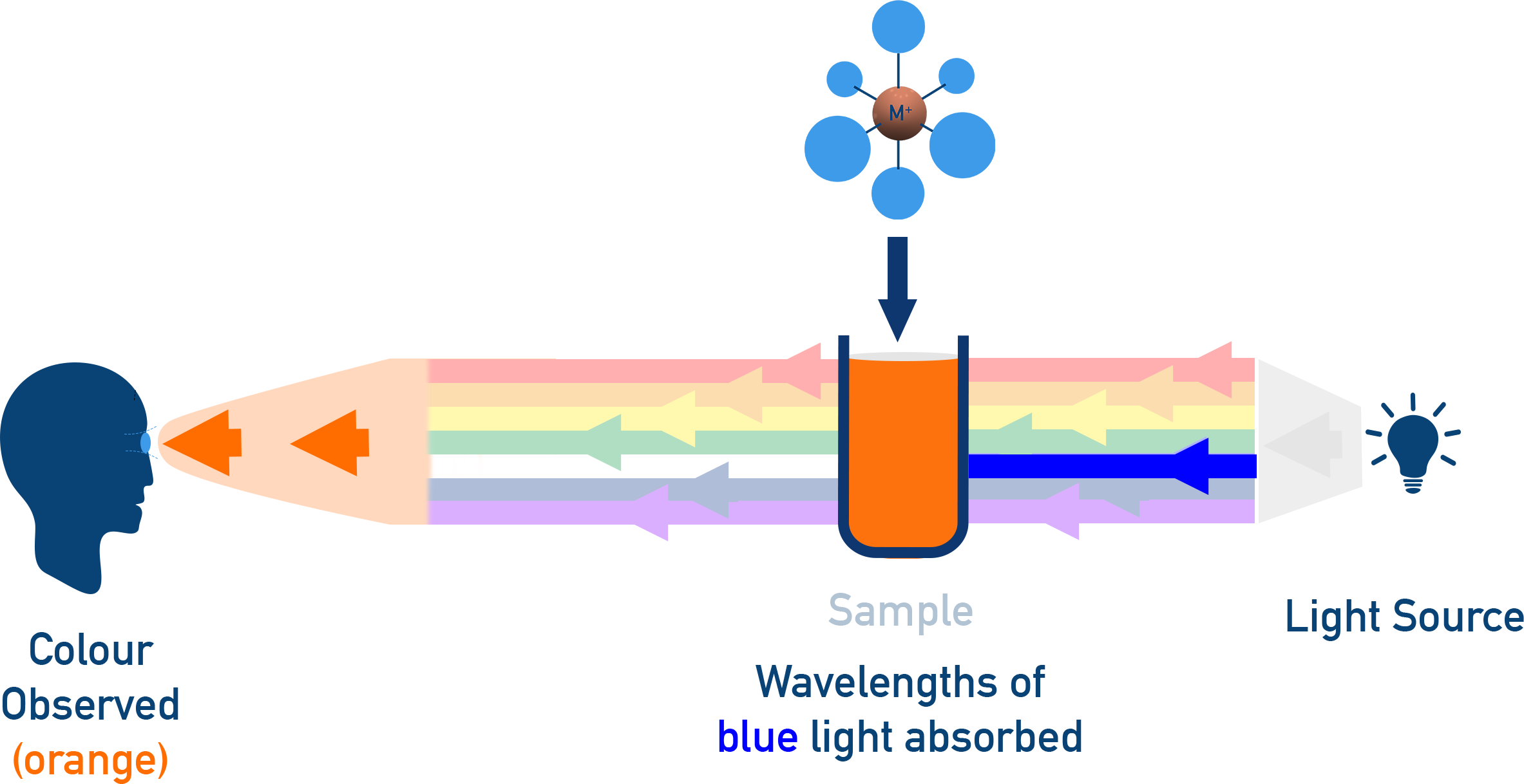
- The colour observed is the complementary colour of the light absorbed.
- Factors affecting the colour of transition metal ions include:
- Oxidation state
- Co-ordination number
- Type of ligand
- Spectroscopy and colorimetry can be used to measure the concentration of coloured ions in solution.
Full Notes
Colour, transition metals and d-orbital splitting have been covered in more detail
here
and
here.
This page is just what you need to know for AQA A-level Chemistry :)
Why Are Transition Metal Ions Coloured?
Transition metals have partially filled d-orbitals.
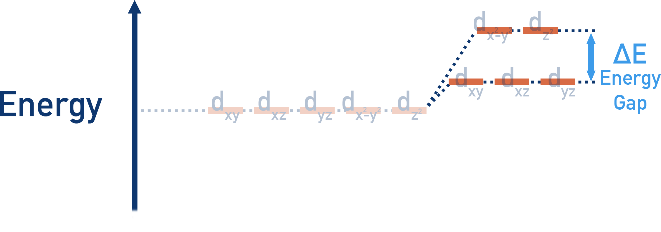
When ligands bond to the metal ion, the outermost d-orbitals split into two energy levels (higher and lower).
This occurs because electrons in the d-orbitals are repelled by electrons from incoming ligands. Due to the shapes of d-orbitals, different orbital shapes experience differing amounts of repulsion – meaning the orbitals get split into different energies. There is an energy gap (ΔE) between the d-orbitals.
Electrons can absorb energy from visible light to move from a lower energy level (ground state) to a higher one (excited state).
The remaining wavelengths of light are transmitted or reflected, giving the solution its observed colour.
The Energy Difference Between d-Orbitals
The energy difference between d-orbitals (ΔE) is given by:
ΔE = hν = hc/λ
-
where:
- h = Planck’s constant (6.63 × 10⁻³⁴ J s)
- ν = frequency of absorbed light (Hz)
- c = speed of light (3.00 × 10⁸ m/s)
- λ = wavelength of absorbed light (m)
If ΔE falls within the visible light spectrum, the compound appears coloured.
This is why transition metals are somewhat unique – the energy gap does fall in the visible light region. Organic molecules, for example, tend to absorb light in the ultraviolet (UV) region, which is why they have no visible colour.
If all wavelengths are absorbed (or none are absorbed), the compound appears colourless.
Factors Affecting the Colour of Transition Metal Complexes
Oxidation State
Different oxidation states cause different energy gaps, changing the colour observed.
Example Fe2+ (pale green); Fe3+ (yellow/brown)
Co-ordination Number
Changing the number of ligands affects d-orbital splitting.
Example [Cu(H2O)6]2+ (blue, co-ordination number 6) → [CuCl4]2− (yellow-green, co-ordination number 4)
Type of Ligand
Different ligands split d-orbitals by different amounts, changing the energy absorbed.
Example [Co(H2O)6]2+ (pink, with H2O ligands) vs [Co(NH3)6]2+ (yellow, with NH3 ligands)
Spectroscopy and Colorimetry
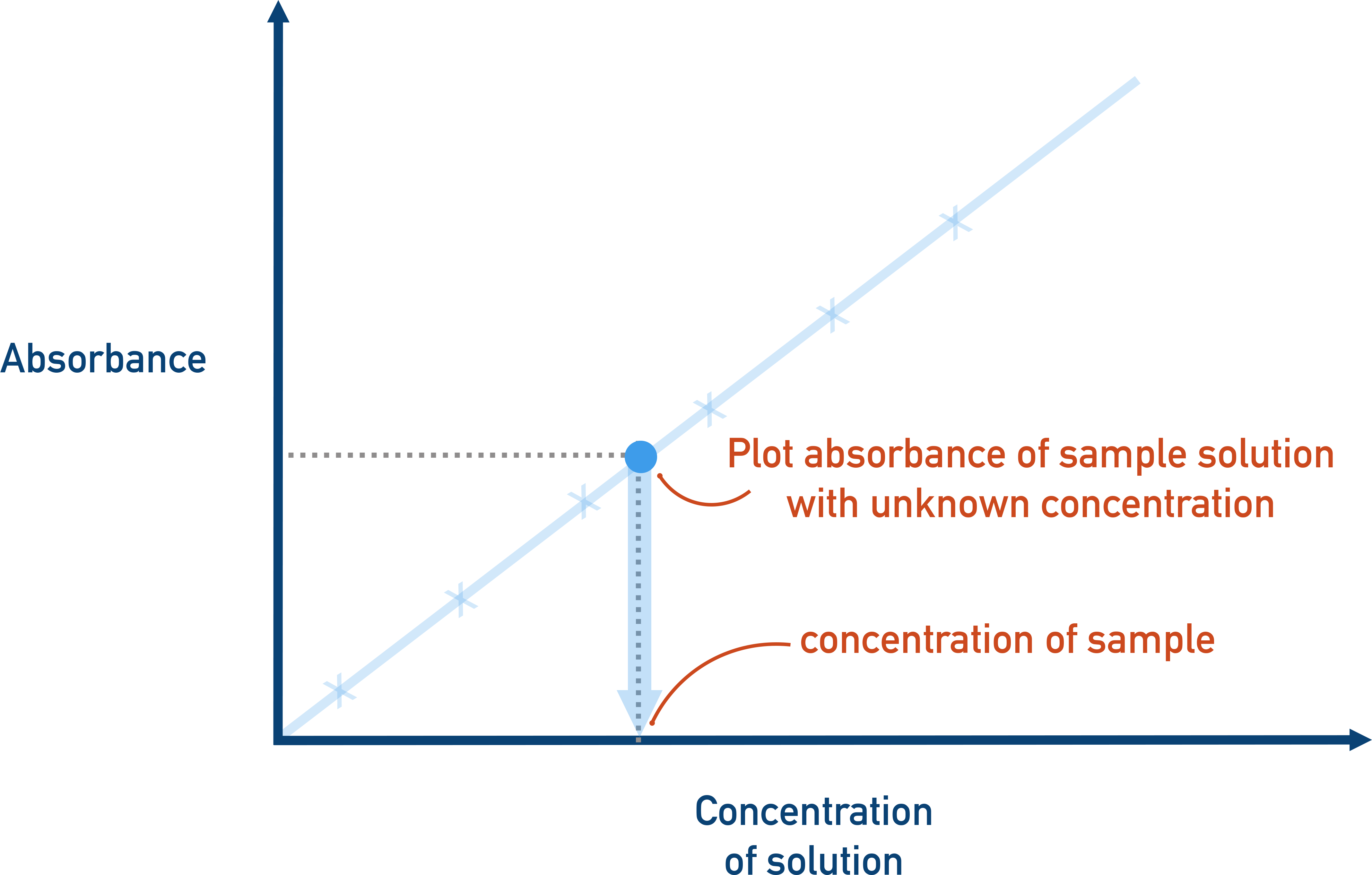
Colorimetry is used to determine the concentration of a coloured solution.
A simple colorimeter measures absorbance of light at a particular wavelength.
Higher absorbance = higher concentration of coloured ions.
How a colorimeter works:
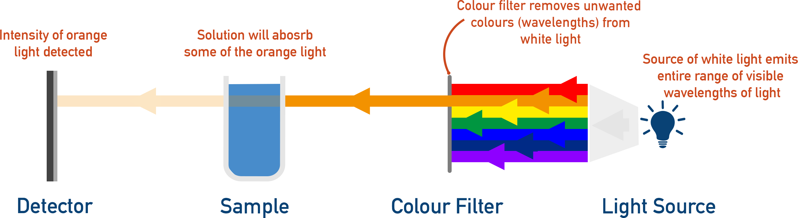
- A filter selects the wavelength of light absorbed most strongly by the solution.
- Light passes through the solution.
- The detector measures how much light is absorbed.
- A calibration curve is used to determine concentration.
Summary Table
| Concept | Key idea | Example / note |
|---|---|---|
| d-orbital splitting | Ligands split d-orbitals into two energy levels; electrons absorb visible light to promote between levels | ΔE within visible region = coloured compound |
| Energy relation | ΔE = hν = hc/λ | Complementary colour observed |
| Oxidation state | Alters ΔE and hence colour | Fe2+ pale green; Fe3+ yellow/brown |
| Co-ordination number | Changing CN changes splitting | [Cu(H2O)6]2+ (blue, CN 6) vs [CuCl4]2− (yellow-green, CN 4) |
| Ligand type | Different ligands split d-orbitals by different amounts | H2O vs NH3 for Co2+ |
| Colorimetry | Absorbance at chosen λ correlates with concentration | Use calibration curve |